Eight things hiding in a drop under the lens

DC·54 Deep Cuts
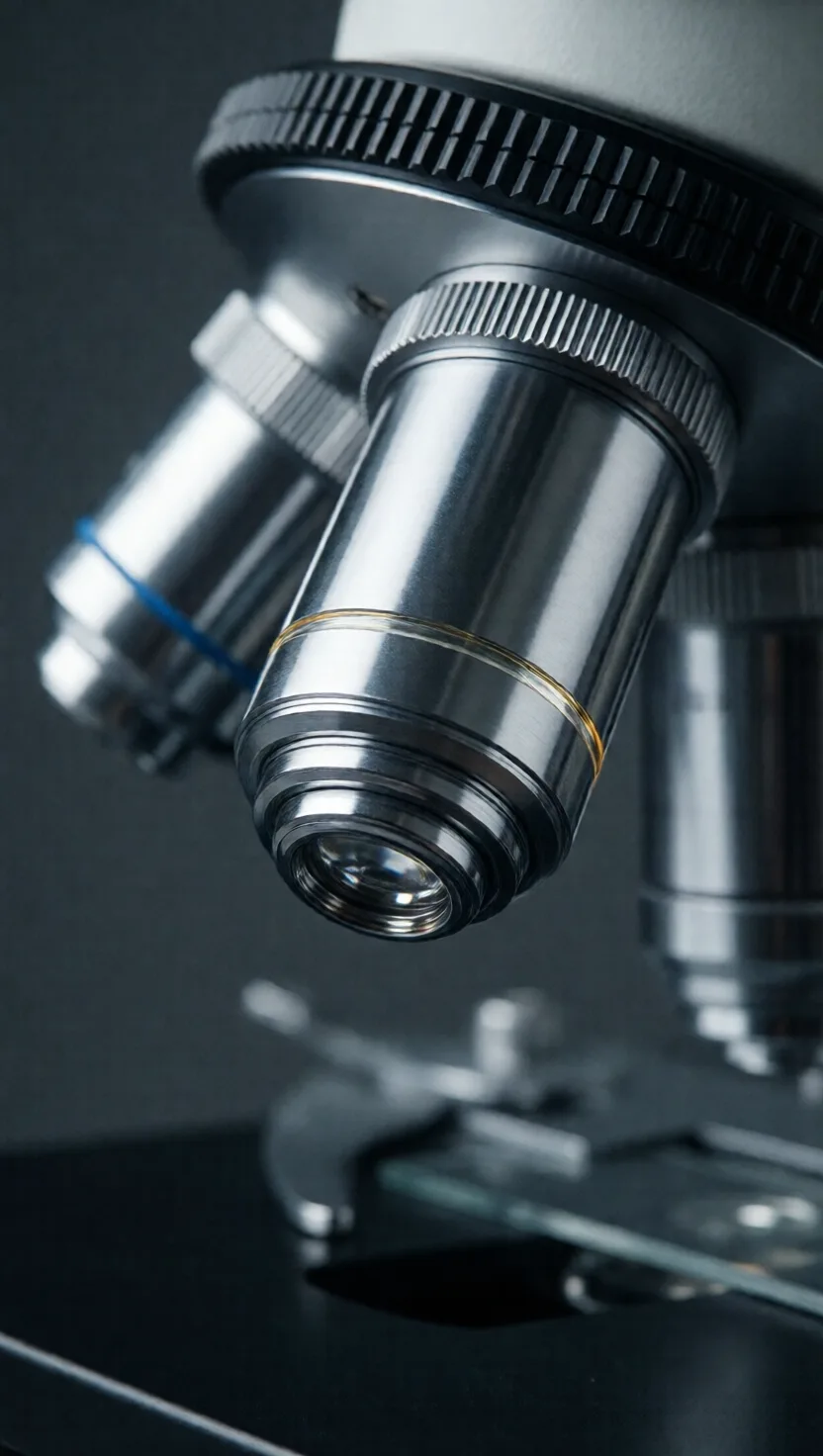
One tiny glass bead beat every microscope for 200 years

One tiny glass bead beat every microscope for 200 years

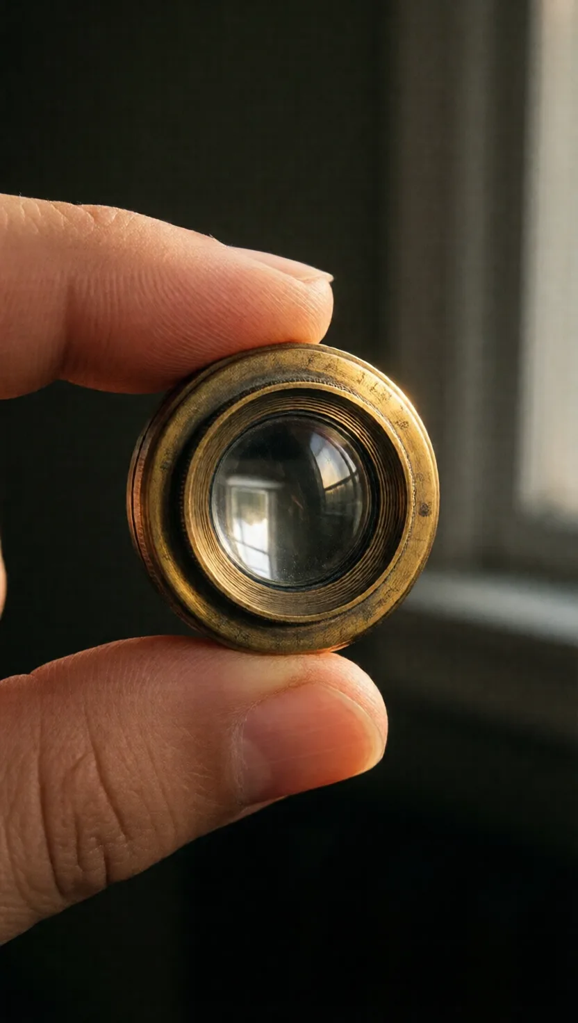
A self-taught Delft draper ground a single grain-sized glass bead into a lens and set it in a brass plate barely the size of a postage stamp. Squinting through it, he saw bacteria and living cells no one knew existed. His simple lens reached about 270x and stayed sharper than the bulky compound microscopes of the day for nearly two centuries, until lens-making finally caught up in the 1800s.
He named the 'cell' after a monk's bare room

He named the 'cell' after a monk's bare room

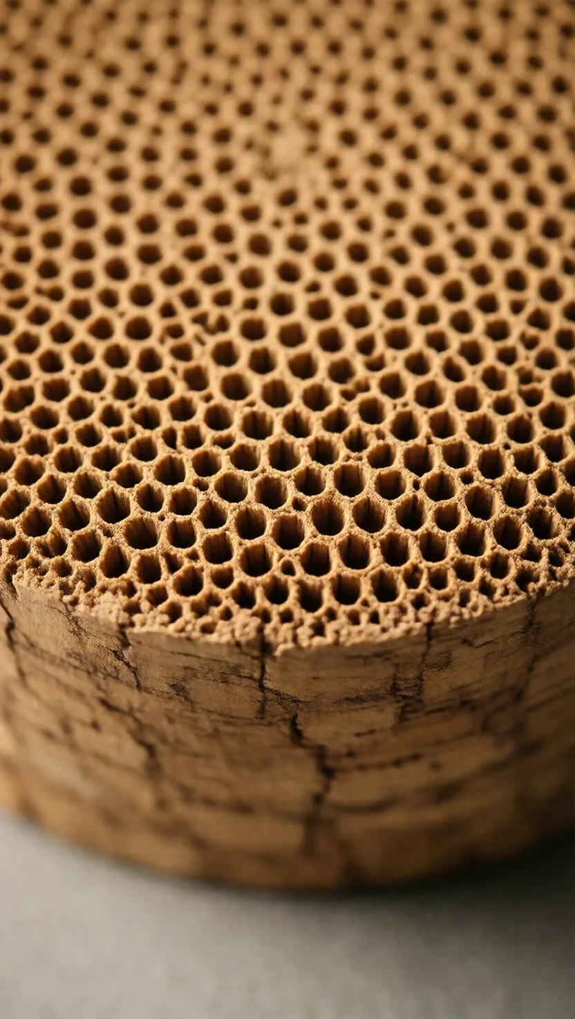
In 1665 a London polymath sliced a wafer of cork, held it under his microscope, and saw a honeycomb of tiny empty boxes. The walls reminded him of the plain little rooms where monks slept, so he borrowed their name: cella, Latin for a small chamber. Every living cell ever since carries that word, first coined for the dead, hollow walls of bark.
Light itself sets the smallest thing a lens can show

Light itself sets the smallest thing a lens can show

In 1873 a German physicist proved that no ordinary light microscope, however perfect its glass, can separate two points closer than about half the wavelength of the light passing through them. For visible light that floor is roughly 200 nanometres, smaller than most bacteria but far larger than a virus. The barrier isn't the lens. It is the wave nature of light itself.
A drop of oil sees sharper than air ever could

A drop of oil sees sharper than air ever could

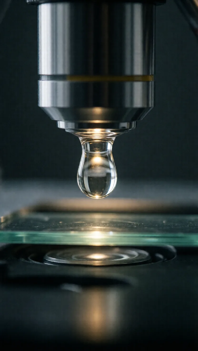
Air bends and scatters the steepest light rays leaving a specimen, so they never reach the lens. Put a drop of special oil between the glass slide and the objective and, because the oil bends light almost exactly like glass does, those rays travel straight in. Matching the oil to the glass lets the lens gather a wider cone of light and resolve detail no dry lens can reach.
Glassy algae are the rulers that test a lens

Glassy algae are the rulers that test a lens

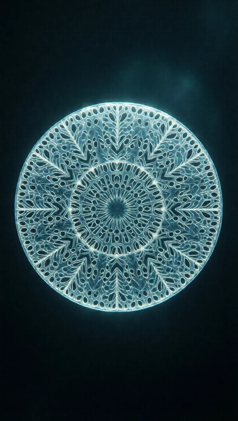
Diatoms are single-celled algae that build intricate shells of glassy silica, pierced with rows of pores spaced just micrometres apart. Those rows are so evenly ruled that microscope makers use them as natural test targets: if your lens can cleanly split the lines of a given species, you know its true resolving power. Some species' stripes sit only a few millionths of a metre apart.
How we finally watched a living cell without killing it

How we finally watched a living cell without killing it

Most cells are nearly transparent, so for decades the only way to see their insides was to stain them with dye, which killed them. In the early 1930s a Dutch physicist realised that a clear cell still subtly delays the light passing through it. His phase-contrast microscope turns those invisible delays into visible brightness, revealing the machinery of a living, unstained cell. It won the 1953 Nobel Prize in Physics.
Swap light for electrons and you can see atoms

Swap light for electrons and you can see atoms

Because light can't resolve anything smaller than about half its wavelength, there is a hard floor to what an optical microscope shows. Electrons, it turns out, behave like waves too, with wavelengths thousands of times shorter than visible light. Fire a focused beam of them at a specimen instead of light and the resolving power leaps a thousandfold, sharp enough to image the lattice of individual atoms.
A second beam of light broke light's own limit

A second beam of light broke light's own limit

For over a century the wavelength of light seemed an unbreakable wall on microscope detail. Around 2000 a physicist added a second, doughnut-shaped laser beam that switches off the glow everywhere except a spot far smaller than the old limit, then scans that spot across the sample. The trick shattered the barrier and let light microscopes see structures just tens of nanometres apart. It earned a 2014 Nobel Prize.
tap →swipe ↑ for depthswipe ↓ to exit